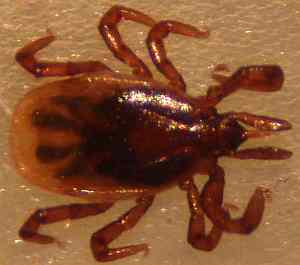
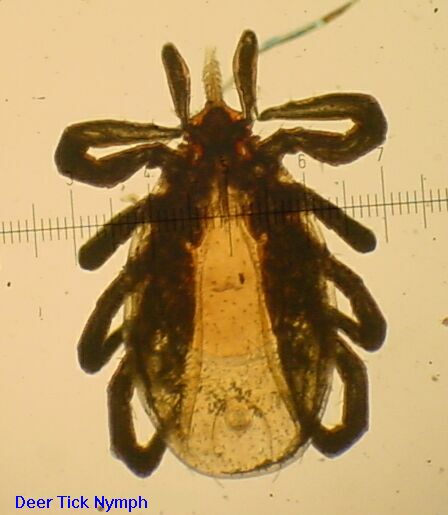
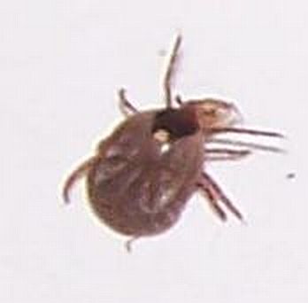
Deer Tick Nymph, Live
© 2002 - 2022 by John Moran, Newtown,CT, USA
Deer Tick Nymph, Live
These tick pictures are an adjunct to the paper Dealing with Deer Ticks provided on this site. Most people are not familiar with deer ticks and cannot tell a deer tick from a dog tick -- the goal here is to help in identification as well as to provide information on how to tell when a tick may be carrying Lyme disease and when it is very unlikely. All you need is a 10 power magnifier to determine whether a deer tick is a larva (6 legs, unlikely to carry Lyme) or a nymph or adult (8 legs, commonly infected with Lyme and/or other tick borne illnesses here in CT) -- read the paper on this site for more info. The numbered scale shown in most of the pictures can be used to judge the exact size but you must be somewhat familiar with the tick type to determine which objective was used to take the picture -- 250/100/25 microns per numbered division (1000 microns=1mm) are produced by the 4/10/40 power objectives respectively. The tick pictures here are available by clicking on the descriptions, this to minimize bandwidth for those who arrive here by accident.
Another site with tick identification pictures.
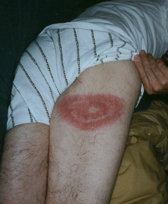
Lyme Disease Rash, I was bitten in the back of the thigh; it took 10 days to develop this classic rash.
Lyme Rash #2, I was bitten several years later on the left arm; small rash this time.
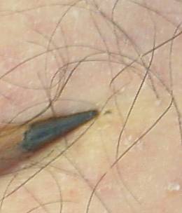
Larva in situ, just beyond pencil point. Small, aren't they?
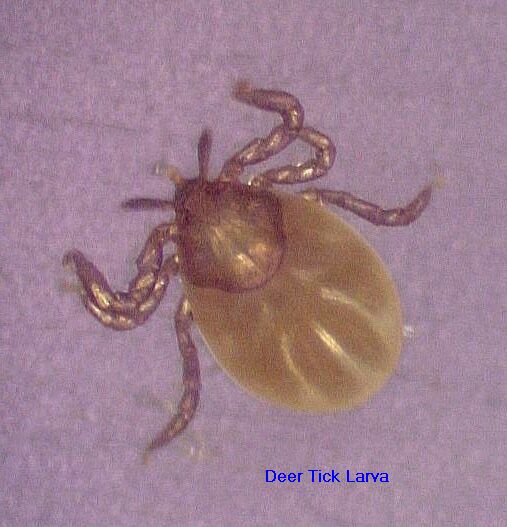
Deer Tick Larva, note 6 legs. This is the same larva shown above, after removal
Deer Tick, lost the left palp when I removed it from my hide
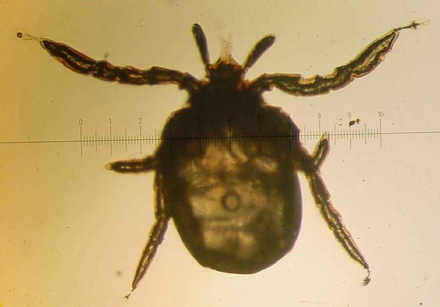
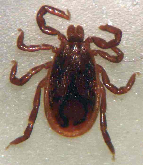
Deer Tick Nymph, note 8 legs. Compare to dog tick below
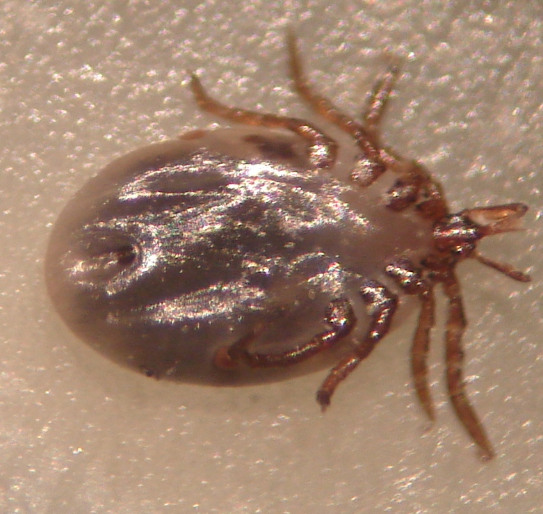
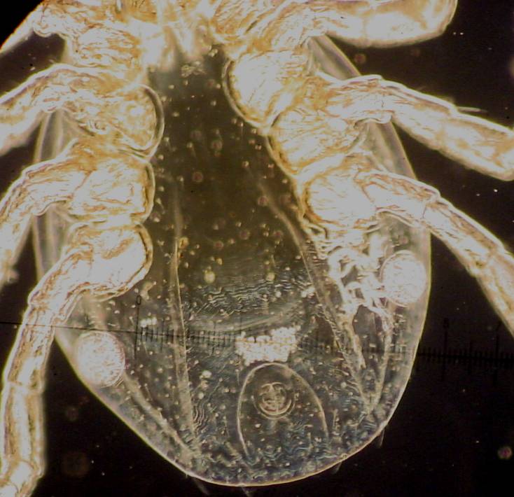
Deer Tick Nymph, bottom view live, Adults have a genital pore mid-way between rearmost legs
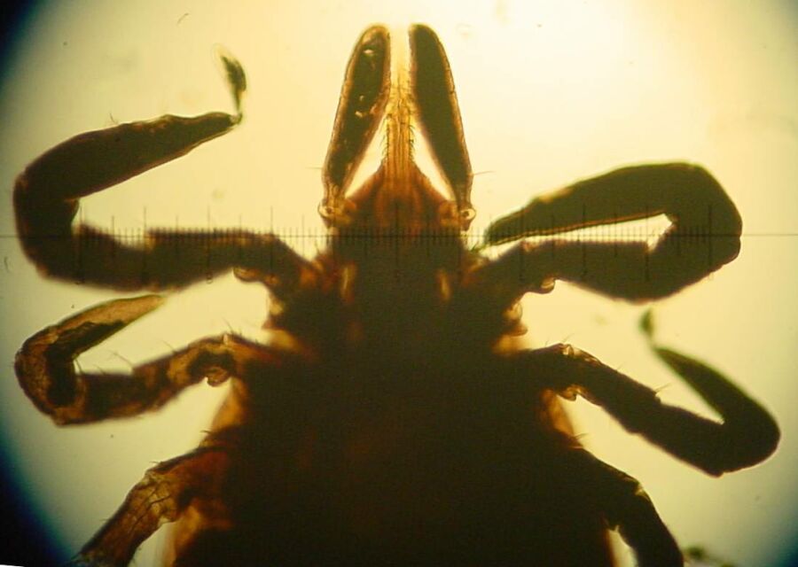
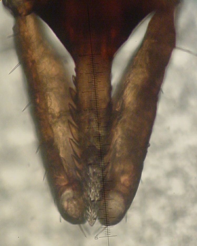
Nymph, business end
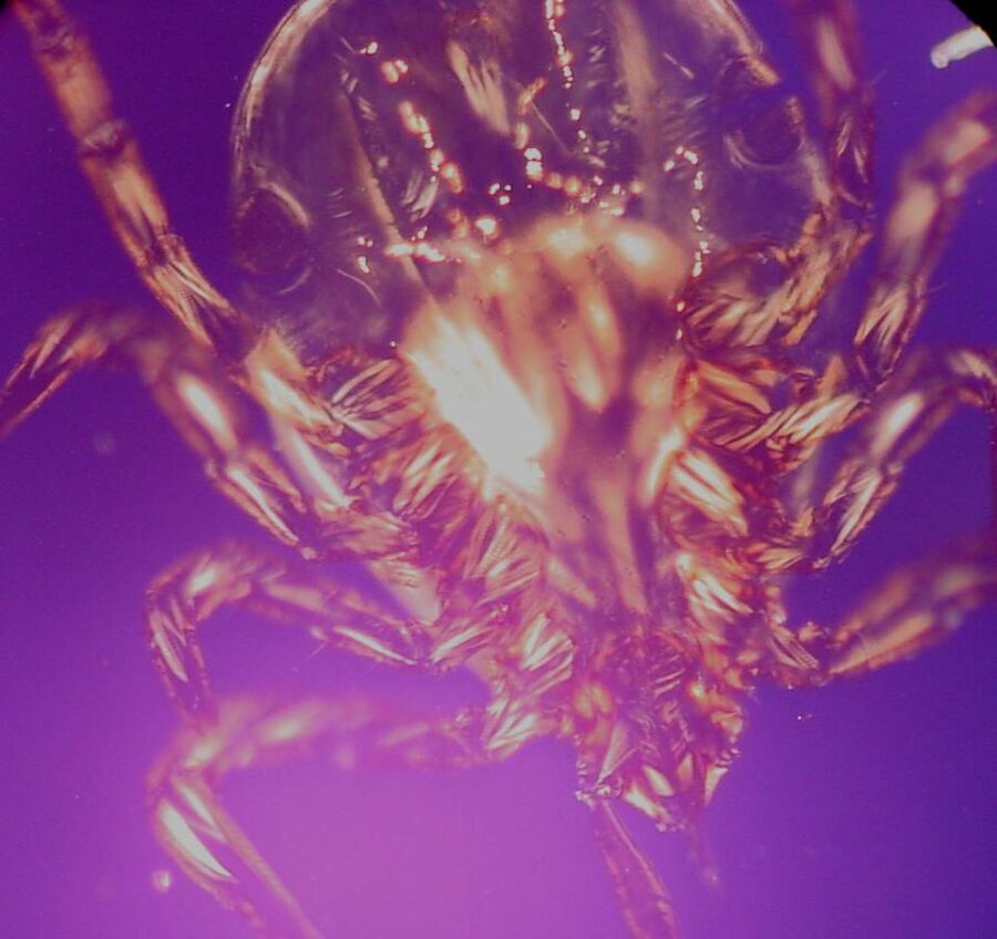
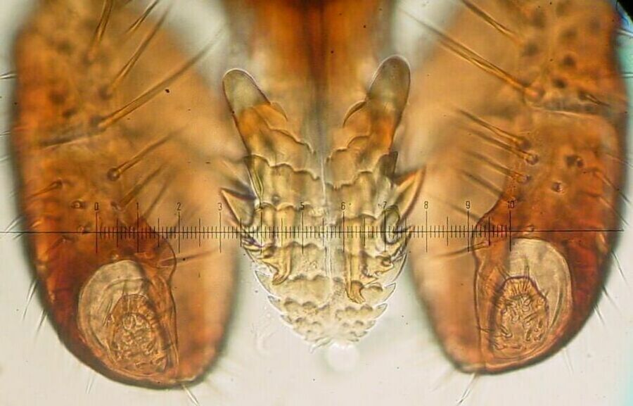
Closeup of hypostome and palps
Another closeup of hypostome and palps
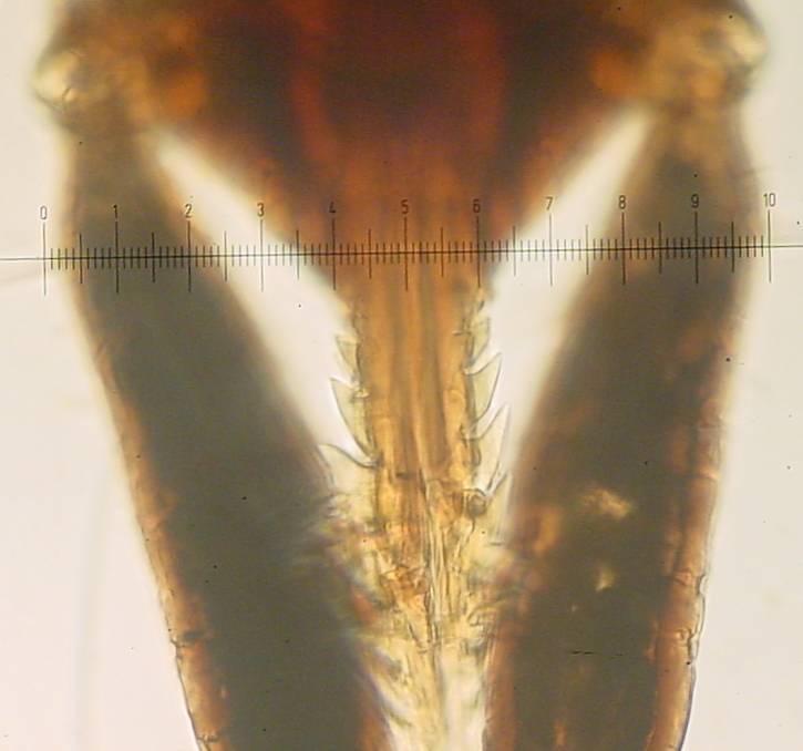
Another view of the hypostome, the holdfast organ with chelicerae partially extended
Even closer -- this is why they're hard to remove
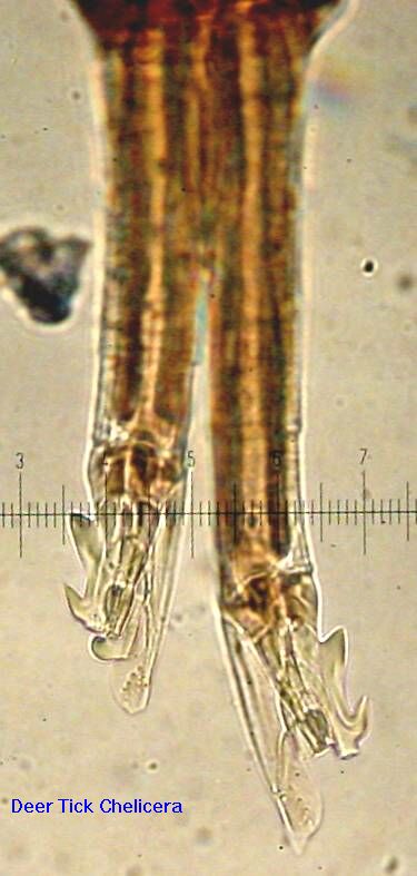
The chelicerae, used to cut an opening for insertion of the hypostome
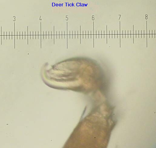
Tick claw, as used to snag a passing host
Larva in questing stance
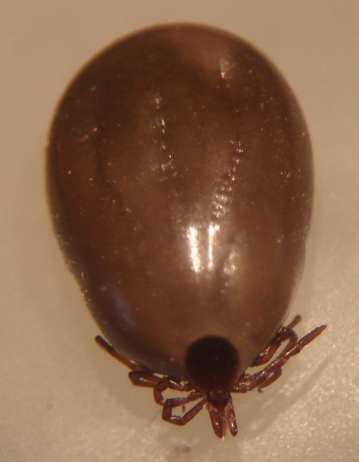
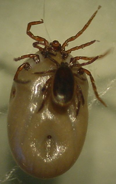
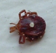
Female deer tick partially engorged, live. Ruler is 1mm per division so it is about 6mm
Female deer tick engorged, top view. The dark brown scutum, which doesn't change size, is about half the body length prior to engorgement.
Pair mating Female's head damaged on removal from our cat
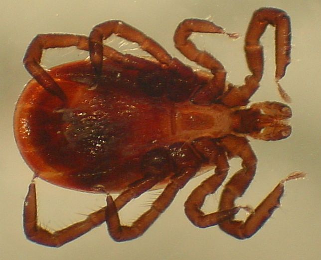
Male from above pair, length is about 2.3mm. Note the short, round palps similar to a dog tick
Male, top view and bottom view
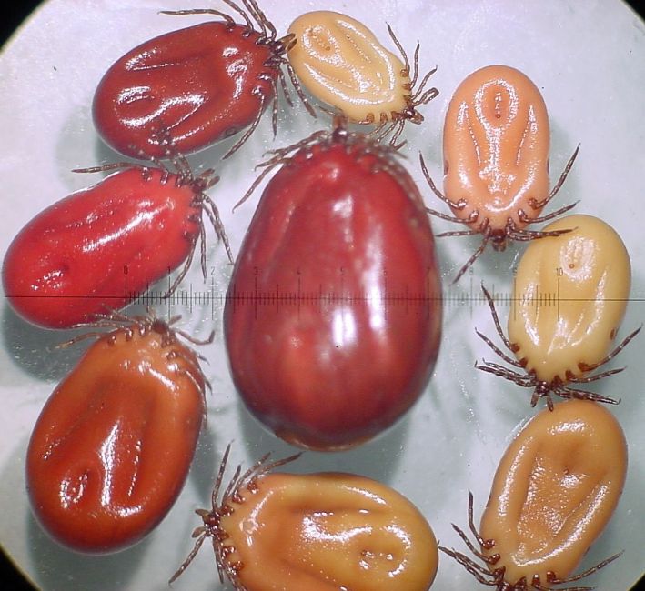
Several Female deer ticks partially engorged, to show color and size variation. Largest is 10mm.
Ticks breathe via round spiracles located aft of the legs
Polarized light shows the tick's muscles
Tick + Tweezer -- Showing why it is difficult to grab a tick by the mouthparts or head (capitulum). The hypostome broke off when this tick was removed from our cat. We remove ticks from people with an X-Acto knife, see this paper for details.
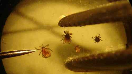
Ticks and a common pin along with forceps for scale. Compare to the theoretical removal method here - makes me wonder if the author at the CDC has ever seen a deer tick. The deer ticks shown are l-r: adult, nymph, and two larvae (mounted on a microscope slide).
The Lone Star tick is similar in size to a deer tick but has a white spot on its back. They are not common in CT (yet) so I have only pictures sent by readers of this site who asked about the tick type.
Lone Star Tick (dead) -- Note the white spot.
Lone Star Tick removed from Sophie the dog.
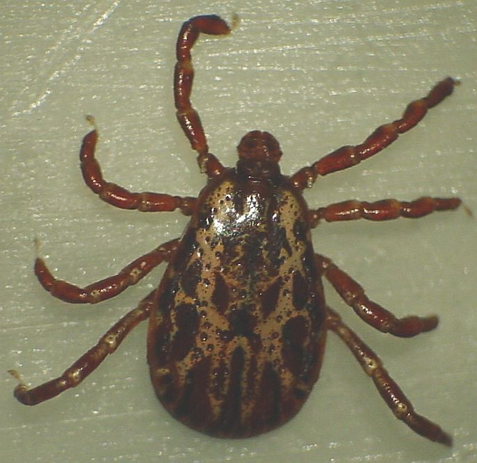
The dog tick larva is similar in size to the deer tick. The dog tick nymph and especially the adult is much larger than the deer tick. While size is the obvious difference between dog and deer ticks note the difference in the shape of the palps and hypostome, see below. The adult American dog ticks (Dermacenter variabilis) have light colored mottling on the back, where the mottling is on the scutum (the shield shape toward the front) in the female and covers the body on the male. Dog ticks have eyes (unlike the deer tick), visible above the second set of legs. The dog tick is also known as the wood tick. The brown dog tick (Rhipicephalus sanguineus) is different in that it lacks mottling and is mainly found in warmer areas than CT - so I don't have pictures.
American Dog tick, adult male, live. Note short, wide palps and the color mottling which is visible to the naked eye
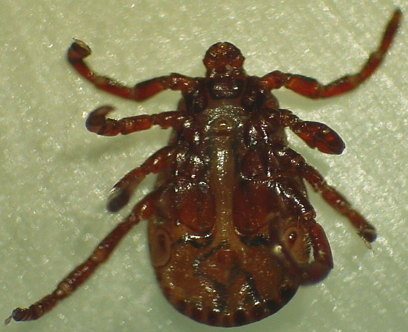
American Dog tick, bottom -- note 11 spots along back edge (festoons). Dog ticks move much faster than deer ticks
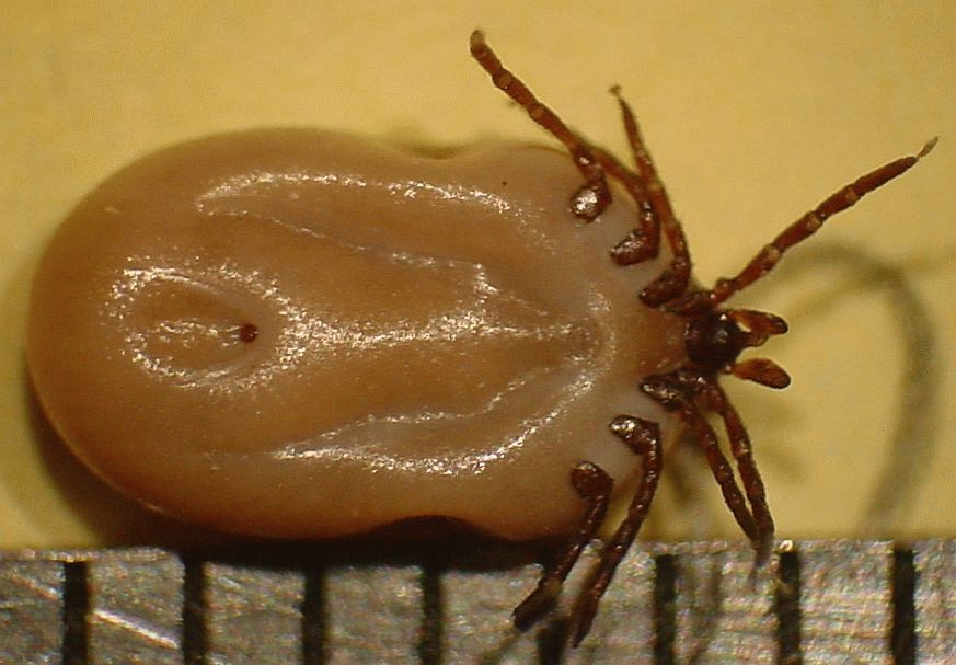
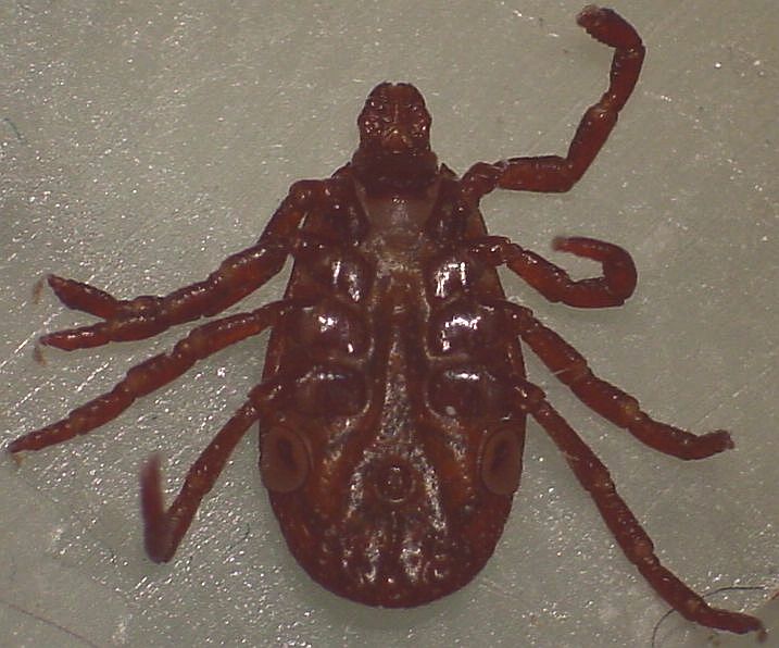
American Dog tick, adult female, live. Note short, wide palps and the color on the shield, visible to the naked eye
American Dog tick, adult female bottom.
Dog tick hypostome and palps. 25u/#div or about 0.001 inch per numbered division. If you look closely the chelicerae are visible through the hypostome.
Deer tick eggs are not commonly found accidentally and are of interest mostly to hard core tick watchers. I found an engorged adult deer tick which dropped off our cat, kept it in a plastic container with a few leaves plus a teaspoon of water, and checked after 10 days. The eggs were in three separate masses, the largest of which is shown below. The eggs appear oily and stick to the cat whisker I use to manipulate samples; grains of sand are also visible stuck to the eggs.
A mass of deer tick eggs. I estimate over 500 in this mass.
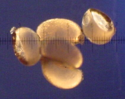
Several eggs, 1/4 mm per numbered division
Eggs magnified to show surface texture
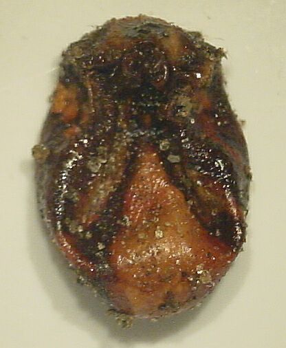
After laying eggs the tick dies, note the shriveled appearance (bottom view).
Additional pictures for the deer tick afficianado. This is a live deer tick nymph on a piece of tape; the tape shows in the background but is necessary to keep the tick from making a getaway.
The nymph, like the eggs, appears oily when incident light is used. 100u/#div
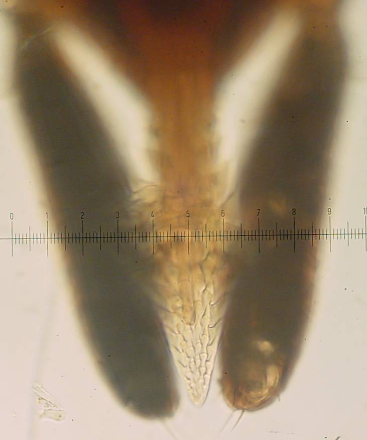
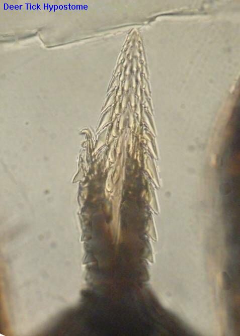
The hypostome and palps, compare to the dog tick hypostome above. 25u/#div
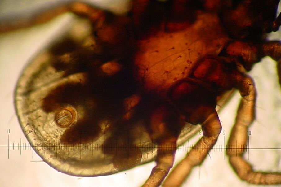
An interesting picture with a number of details. The round ornate structure aft of the rear leg is the spiracle, used for breathing. The holes visible on the spiracle connect to air tubes; these air tubes and their surrounding blood vessels are visible as a dark mass adjacent to the spiracle. The round circle with a longitudinal line in the aft center of the tick is the anus with adjacent dark internal structures. In the center of the tick the redish brown thorax shows several short hairs plus a long blood vessel running lengthwise plus a few blurry branches from the main vessel. If this were an adult tick there would be a genital pore visible as a lateral line between the rear set of legs. This nymph measured 1.4mm overall, 1mm from the base of the neck to the rear of the body - the scale shown is 100u = 0.1mm per numbered division = approximately 0.004 inch, so the legs are about the thickness of a human hair.
I was bitten by this female tick of unknown type recently. The legs are not dark like a normal deer tick but the palps are long and narrow, typical of a deer tick. The size is on the large end of the deer tick range but small for a dog tick. Overall, it appears to be of genus Ixodes but not scapularis species, i.e. it doesn't seem to be a common deer tick but is likely a close relative, possibly I.ricinis. My references for identification are PracticalScience and GreenValley Pest Control's World of Pests; if you have come across more complete references on the net, please advise me.
There is now a site in the UK dedicated to collecting and identifying ticks, worth a visit just to see the number of different ticks found there.
If you have a comment on my site or its contents, a question about deer ticks,
click here. For tick questions, please include your location.
All pictures and text on this site are copyright by John Moran. In some cases I allow others to use my pictures for educational purposes. You must write to me and obtain written permission to use my pictures and this permission does not allow you to grant others permission to use my pictures -- copyright remains with me. Each of my pictures, when included on another site must be clickable and link back to the page on my site where it is found; view the source for the first picture on this page for an example.